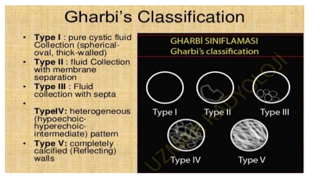
Gharbi Classification Of Hydatid Cyst : 1 - Cystic lesion with daughter lesions.. Calcifications occur in the pericyst; Doppler ultrasonography is indicated to show the reports of hydatid cyst with vascular axes (portal vein, hepatic veins, and inferior vena cava). Gharbi classification of hydatid cysts type description i pure fluid collection ii fluid collection with a detached membrane iii fluid collection with multiple septa and/or daughter cysts iv hyperechoic with high internal echoes v cyst with reflecting. Calcified or partially calcified lesion (inactive cyst) However, in the types i and iv, we have to consider differential diagnosis.
Ce3 are cysts entering a transitional stage where the integrity of the cyst has been compromised either by the host or by chemotherapy. The ultrasonographic (us) appearance of hydatid cysts may vary. Type iii hydatid cysts are those with fluid collection and septa. Proposed a subdivision of the hydatid cysts into five types: The host is at the origin of the image of pericystic wall.
Table 3 From Hydatid Cyst Of The Kidney Diagnosis And Treatment Semantic Scholar from d3i71xaburhd42.cloudfront.net Type ii is purely cystic plus hydatid sand; Yasawy mi, mohammed ae, bassam s, karawi ma, shariq s. The cyst wall usually manifests as double echogenic lines separated by a hypoechogenic layer (, 6). 1, 39 direct rupture may cause massive intra‐abdominal hemorrhage 40 or biliary peritonitis. The host is at the origin of the image of pericystic wall. Cystic lesion with daughter lesions. Usg appearance of hydatid cyst. There was only one case of type iv and no cases of type v.
Usg appearance of hydatid cyst.
Discover (and save!) your own pins on pinterest Touch device users can explore by touch or with swipe gestures. Treatment of gharbi type iii hydatid cysts is still controversial. There was only one case of type iv and no cases of type v. Type iii hydatid cysts are those with fluid collection and septa. The initial classification by gharbi et al and the world health organization classification are the most commonly preferred. The cysts should be larger than 5 cm in diameter and type i or ii according to the gharbi ultrasound classification of liver cysts (ie, type i is purely cystic; It is also helpful for the classification of hydatid cysts. The ultrasonographic (us) appearance of hydatid cysts may vary. The cyst wall usually manifests as double echogenic lines separated by a hypoechogenic layer (, 6). The host is at the origin of the image of pericystic wall. This classification was proposed by the who in 2001 and, at the time of writing (july 2016), remains the most widely used classification for hepatic hydatid cysts. All cysts were classified as per the gharbi's classification.
The liver segments were grouped as near to the hilum (segments i, iii, ivb, v, and vi) and remotely distant (segment ii, iva, vii, and viii) with modification of classification by dziri et al. Left renal hydatid cyst with floating membranes following rupture of the cyst into urinary tract. 1, 39 direct rupture may cause massive intra‐abdominal hemorrhage 40 or biliary peritonitis. Liver hydatidosis is characterized by progressive growth of the hydatid cyst, which in its mature form is a fluid filled cavity, delimited by an external dense host fibrous reaction (pericyst) and two internal parasite derived layers (endocyst). The initial classification by gharbi et al and the world health organization classification are the most commonly preferred.
Mechanical Suction Through Wide Bore Catheters For Nonsurgical Management Of Gharbi Type Iii Hepatic Hydatid Cysts from www.tropicalgastro.com Discover (and save!) your own pins on pinterest It is also helpful for the classification of hydatid cysts. The liver segments were grouped as near to the hilum (segments i, iii, ivb, v, and vi) and remotely distant (segment ii, iva, vii, and viii) with modification of classification by dziri et al. A catheterization technique was performed but hypertonic saline and alcohol were not given into the cavity due to cystobiliary leakage. Fluid collection with a detached membrane table (1) : The cyst wall usually manifests as double echogenic lines separated by a hypoechogenic layer (, 6). The gharbi ultrasound classification consists of five stages 4: And type iv has peripheral or diffuse distribution of coarse echoes in a complex.
Gharbi classification of hydatid cysts type description i pure fluid collection ii fluid collection with a detached membrane iii fluid collection with multiple septa and/or daughter cysts iv hyperechoic with high internal echoes v cyst with reflecting.
The host is at the origin of the image of pericystic wall. Type iii has the membrane undulating in the cystic cavity; It is also helpful for the classification of hydatid cysts. There was only one case of type iv and no cases of type v. Type iii hydatid cysts are those with fluid collection and septa. And type iv has peripheral or diffuse distribution of coarse echoes in a complex. Mechanical suction through wide bore catheters for nonsurgical management of gharbi type iii hepatic hydatid cysts. Yasawy mi, mohammed ae, bassam s, karawi ma, shariq s. Ce3 are cysts entering a transitional stage where the integrity of the cyst has been compromised either by the host or by chemotherapy. Through a sonographic evaluation, gharbi et al. The liver segments were grouped as near to the hilum (segments i, iii, ivb, v, and vi) and remotely distant (segment ii, iva, vii, and viii) with modification of classification by dziri et al. There are several classification schemes for liver hydatid cysts based on their ultrasound appearances; The initial classification by gharbi et al and the world health organization classification are the most commonly preferred.
Usg appearance of hydatid cyst. The gharbi ultrasound classification consists of five stages 4: The initial classification by gharbi et al and the world health organization classification are the most commonly preferred. Through a sonographic evaluation, gharbi et al. There are several classification schemes for liver hydatid cysts based on their ultrasound appearances;
Management Of Hydatid Cyst And Osteoid Osteoma from image.slidesharecdn.com Type iii has the membrane undulating in the cystic cavity; Discover (and save!) your own pins on pinterest A nonpyogenic infective etiology of liver lesion with multilocular cysts within, an imaging differential for melioid liver abscess, is a hydatid cyst with multiple daughter vesicles (world health. And type iv has peripheral or diffuse distribution of coarse echoes in a complex. Liver hydatidosis is characterized by progressive growth of the hydatid cyst, which in its mature form is a fluid filled cavity, delimited by an external dense host fibrous reaction (pericyst) and two internal parasite derived layers (endocyst). Calcified or partially calcified lesion (inactive cyst) Proposed a subdivision of the hydatid cysts into five types: And type iv has peripheral or diffuse distribution of coarse echoes in a complex.
Liver hydatidosis is characterized by progressive growth of the hydatid cyst, which in its mature form is a fluid filled cavity, delimited by an external dense host fibrous reaction (pericyst) and two internal parasite derived layers (endocyst).
The classification proposed by gharbi et al., for liver hydatid disease based. Cystic lesion with daughter lesions. Type iii has the membrane undulating in the cystic cavity; The cysts should be larger than 5 cm in diameter and type i or ii according to the gharbi ultrasound classification of liver cysts (ie, type i is purely cystic; The hydatid cyst grows slowly and remains asysmptomatic for many years. Two of the lesions were type 1, one was type 2, two were type 3 and two were type 4 hydatic cyst according to gharbi classification. This classification was proposed by the who in 2001 and, at the time of writing (july 2016), remains the most widely used classification for hepatic hydatid cysts. A nonpyogenic infective etiology of liver lesion with multilocular cysts within, an imaging differential for melioid liver abscess, is a hydatid cyst with multiple daughter vesicles (world health. Hydatid cyst of the liver is the most common clinical presentation of echinococcus granulosus. Left renal hydatid cyst with floating membranes following rupture of the cyst into urinary tract. 41 the incidence of hcs rupturing into the peritoneal. Cyst types remains unchanged from gharbi's classification and the types are categorized into active, transitional, and inactive stages. According to gharbi's classification, three cases (21.4 %) of the unusually located hydatid cysts were type i, two (14.3 %) type ii, and eight (57.1 %) type iii.


0 Komentar